
Über die Klinik
Eine familiär geführte Zahnklinik in Sopron, aufgebaut um ein Instrument: das Mikroskop.
Harajda Dent ist eine Zahnklinik in Sopron, Ungarn, und behandelt seit mehr als dreißig Jahren am Várkerület. Die Haltung ist über die ganze Zeit dieselbe geblieben: sorgfältig untersuchen, verständlich erklären und ohne Eile behandeln.
Unsere Haltung
Wie wir mit Patientinnen und Patienten arbeiten.
In unserer Klinik empfangen wir Sie und Ihre Familie in einer freundlichen Umgebung, in der nichts gehetzt wird. Unser erstes Anliegen ist einfach: die Angst nehmen, mit der viele ankommen, ihre Beschwerden schmerzfrei behandeln und eine Beziehung aufbauen, die auf Vertrauen beruht.
Mit mehr als dreißig Jahren Berufserfahrung und moderner Ausstattung möchten wir die zeitgemäßesten zahnerhaltenden und zahnersetzenden Lösungen anbieten, die verfügbar sind, statt auf die Möglichkeit auszuweichen, die am meisten Zahn kostet.
Wir möchten, dass unsere Patientinnen und Patienten die Behandlung, die wir für nötig halten, mit Zuversicht angehen, und ebenso wichtig: dass sie nach deren Abschluss die Gewohnheit regelmäßiger Kontrollen entwickeln.
Wenn unsere Klinik zu Ihnen passt, freuen wir uns auf Sie. Termine werden werktags zwischen 9:00 und 17:00 Uhr telefonisch oder per E-Mail vereinbart. Eine Warteliste für Spontanbesuche gibt es nicht, der erste Schritt ist also immer die Terminvereinbarung.
Räume und Ausstattung
Drei Behandlungszimmer, und die Geräte, die bestimmen, was wir sehen.
Die Klinik arbeitet aus drei Behandlungszimmern, und Röntgen- und 3D-Aufnahmen entstehen im Haus, eine Diagnose wartet also nicht auf eine Überweisung. Nichts davon ist in Westeuropa außergewöhnlich. In Sopron ist es noch ungewöhnlich genug, um es zu erklären, denn es verändert, was eine Zahnärztin sehen kann, und damit auch, was sie erhalten kann.
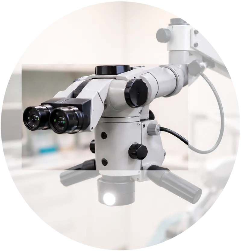
Das Operationsmikroskop für Wurzelkanalbehandlungen und Rekonstruktionen Zahnärztliches Operationsmikroskop
Wurzelkanalbehandlungen und größere Rekonstruktionen werden mit bis zu 25-facher Vergrößerung durchgeführt. Damit lassen sich Kanäle auffinden, die für das Auge und im Röntgenbild unsichtbar sind, Instrumente bergen, die bei einer früheren Behandlung anderswo abgebrochen sind, und für dasselbe Ergebnis weniger gesunde Zahnsubstanz entfernen.
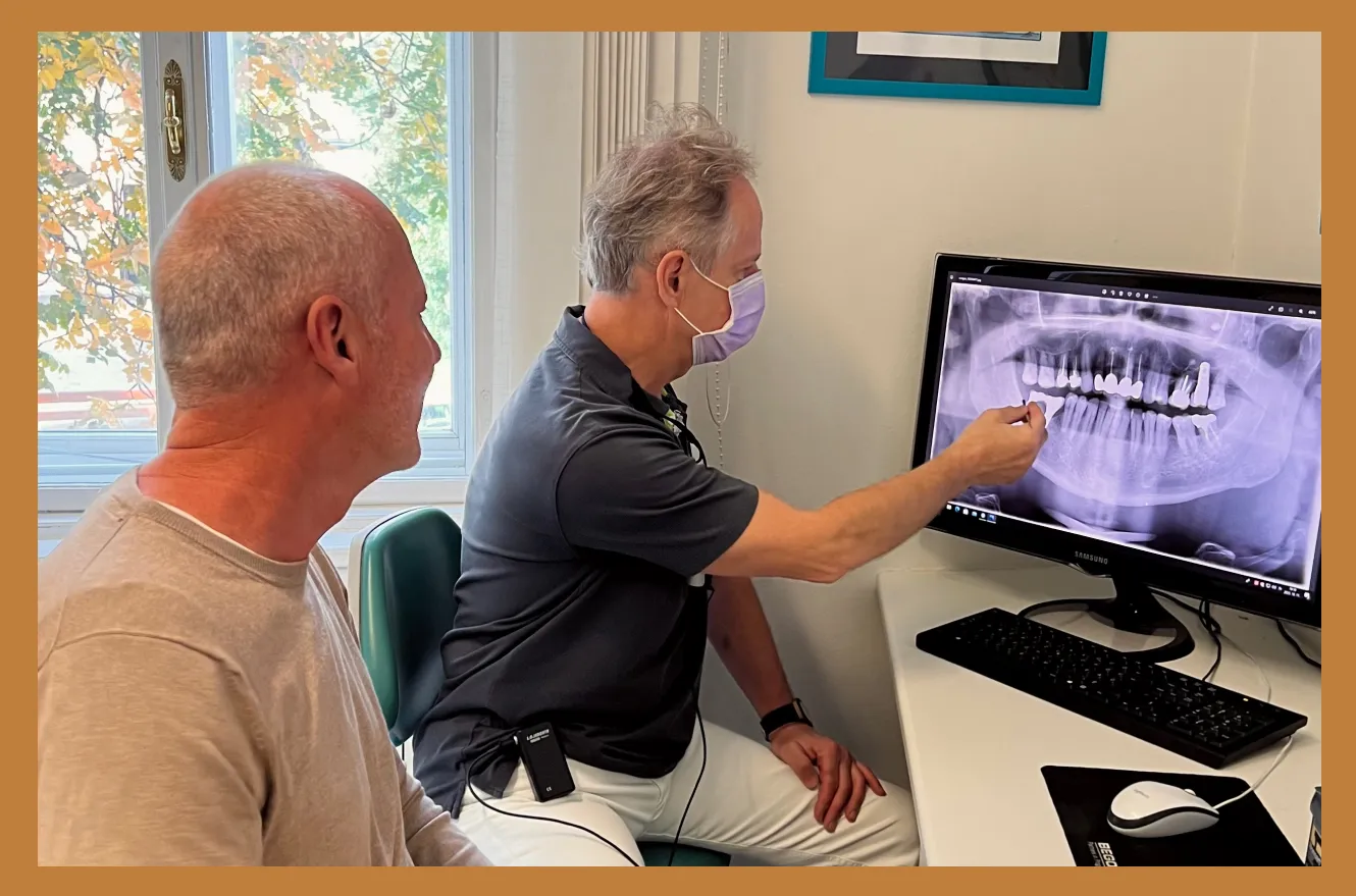
Ein digitaler Scan wird vor der Behandlungsplanung mit einer Patientin durchgesehen Digitale Abformung
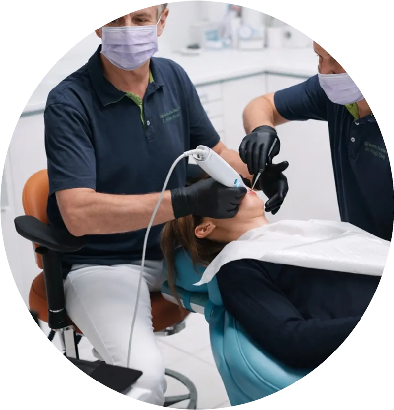
Ein Intraoralscanner baut ein 3D-Modell des Mundes direkt am Bildschirm auf. Keine Abformlöffel, keine Abformmasse, kein Würgereiz und nichts, was wiederholt werden muss, wenn der erste Versuch unsauber war. Der Scan ist zugleich die Grundlage für die Planung von Kronen, Brücken und Implantatarbeiten.
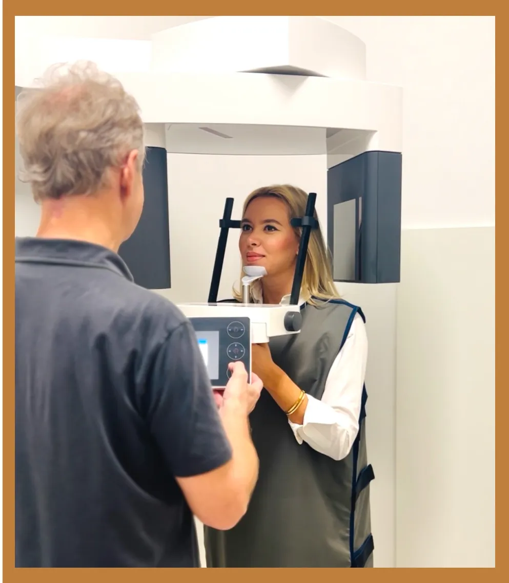
Panorama- und 3D-Aufnahmen, in der Klinik erstellt Röntgen und 3D-Aufnahmen im Haus
Ein Panoramaröntgen zeigt den ganzen Kiefer in einem flachen Bild: welcher Zahn wo steht, wie die Wurzeln liegen, wo eine Entzündung begonnen hat. Eine 3D-Aufnahme, in der Zahnmedizin DVT genannt, liefert statt eines Bildes ein Volumen, und danach werden chirurgische Eingriffe geplant: wie viel Knochen einem Implantat zur Verfügung steht, wo genau ein Weisheitszahn am Nervkanal liegt und was eine überlagernde Wurzel im flachen Bild verdeckt hat. Beides entsteht in der Klinik.
Die Klinik
Geführt von Dr. Harajda Miklós.
Die bisherige Website nennt überhaupt keine Behandelnden, und diese Lücke schließen wir hier zuerst. Dieser Abschnitt wächst, sobald das übrige zahnärztliche Team feststeht.
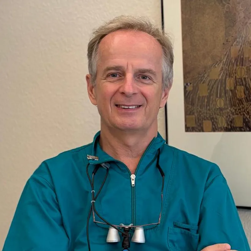
Dr. Harajda Miklós
Leitender Zahnarzt
Dr. Harajda führt eine Klinik, hinter der mehr als dreißig Jahre klinische Erfahrung stehen, mit einem Schwerpunkt auf der Wurzelkanalbehandlung unter dem Mikroskop und auf zahnerhaltender Arbeit: einen Zahn so behandeln, dass er bleiben kann, statt ihn zu ersetzen.
Fachzahnarzt für Zahnheilkunde · Implantologe
Weitere Teammitglieder werden hier vorgestellt, sobald die Klinik ihre Namen, Aufgaben und Qualifikationen bestätigen kann.
Seite zuletzt aktualisiert: August 2026
Die Klinik kennenlernen
Sehen Sie, wie eine Untersuchung ohne Eile aussieht.
Die erste Beratung ist kostenlos und verpflichtet zu nichts. Sagen Sie uns, was Sie stört, und wir gehen von dort aus weiter.
Oder rufen Sie uns an+36 (99) 50 60 90